-
We detected your language preference as English. Would you like to switch to the English version for a better experience?
Switch to English
Stay here


Named after a German chemist, Giemsa stain employs a combination of stains to demonstrate the presence of parasites in diseases like malaria. It belongs to a group of stains known as Romanowsky stains, composed of a mixture of methylene blue, azure, and eosin Y. These neutral stains are applied on air-dried, methanol-fixed glass slides. Romanowsky stains are utilized in cytological differentiation, pathological examination of samples such as blood and bone marrow, and showcasing parasites (e.g., malaria).
Initially used to detect malaria parasites in blood smears, Giemsa stain is now a routine in histology for examining blood smears. It remains one of the most commonly used microscopic stains, extensively employed in hematology for blood and bone marrow specimens, bacteriology, clinical cytology specimens, tissue biopsies, and tumor samples.
Giemsa stain is a differential stain comprising eosin dye, methylene blue, and azure. It specifically binds to phosphate groups of DNA and interacts in regions with high adenine-thymine content, characteristic of DNA.
Azure and eosin are acidic dyes, imparting varying degrees of staining on cellular components such as cytoplasm and granules. Methylene blue, an alkaline dye, stains acidic components of cells, particularly nuclei. Besides its role as a cellular dye, methanol is also used to "fix" images. This fixation allows cells to adhere to glass slides, preventing any further cellular changes.
(1) Wright-Giemsa stain is commonly used to display cellular components in peripheral blood and bone marrow smears. Giemsa stain is utilized for obtaining differential white cell counts. It is also used to differentiate nuclear and cytoplasmic morphology of various blood cells like platelets, red cells, and white cells.
(2) In microbiology, Giemsa stain is employed for staining inclusion bodies in Chlamydia trachomatis, Borrelia burgdorferi (without Warthin-Starry stain), and Yersinia pestis. Giemsa stain is also used for staining capsular tissue of Cryptococcus neoformans, Pneumocystis jirovecii, Klebsiella granulomatis (formerly Calymmatobacterium granulomatis), and occasionally bacterial capsules.
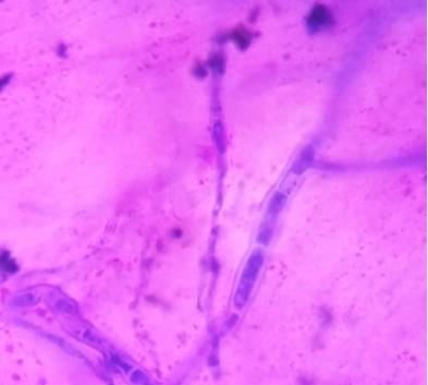
In microbiology, this stain is most commonly used in parasitology to detect intracellular (malaria, Babesia) and extracellular (trypanosomes, microfilariae) parasites. It is also used to detect Leishmania donovani or Trypanosoma cruzi intracellular amastigotes.
(3) Cytogenetics also utilizes this stain to stain chromosomes and identify chromosomal aberrations. It is commonly used in G-banding (Giemsa-Banding). The principle involves staining chromosomes with Giemsa dye after treatment with trypsin, displaying distinctive banding patterns for the 24 human chromosomes.
Giemsa stain is crucial for observing chromosomes, particularly in detecting cytomegalovirus infections where typical inclusions are seen as "owl's eye" viral inclusions, as depicted below:

Red blood cells: Pale purple
Neutrophils: Red-purple
Eosinophils: Purple nuclei with red-orange granules
Basophils: Purple nuclei with blue coarse granules
Lymphocytes: Deep blue nuclei
Platelets: Violet granules
Host cell nuclei: Deep purple
White cell nuclei: Deep purple
Host cell cytoplasm: Light blue
White cell cytoplasm: Light blue or grey-blue
Melanin granules: Black-green
Bacteria: Light blue or dark blue
Spirochetes: Pale purple
Malaria parasite: Red or pink nuclei with blue cytoplasm
Monocytes: Pink cytoplasm
To achieve consistent and reliable Giemsa stain results, several factors need attention. Firstly, the quality of the blood smear itself is crucial. Improperly prepared slides can lead to uneven staining or poor cell morphology. Additionally, the staining solution and its application significantly impact results. Aging or improper dilution of the stain can result in inadequate staining or excessive background staining. Factors like fixation time and rinsing procedures also influence the final product.
Techniques for achieving consistent Giemsa staining include meticulous preparation and adherence to standardized protocols. Blood smears should be air-dried or methanol-fixed as per established guidelines. Freshly prepared Giemsa staining working dilution should be buffered to a specific pH each time. Standardized staining times and rinsing steps ensure reproducibility. Additionally, some protocols incorporate staining coplin jars with agitation mechanisms to promote even staining distribution.
Common staining issues often stem from specific factors. Light staining may result from insufficient staining concentration or duration. Conversely, deep staining may indicate overly concentrated stain or excessive staining duration. Uneven staining may result from inadequate rinsing or uneven application of the stain. By identifying issues and adjusting corresponding factors, Giemsa staining can be optimized to produce clear, informative microscopic images.
The giemsa stain principle technology lies in its specific binding of stains to different components within cells and tissues, enabling clear visualization of cellular structures. This simple yet effective staining method plays a crucial role in biological research and medical diagnostics, providing scientists with an important tool to observe and analyze internal structures of organisms.
[1] https://en.wikipedia.org/wiki/Giemsa_stain/
[2] https://www.creative-bioarray.com/support/giemsa-staining-protocol.htm
[3] https://microbenotes.com/giemsa-stain-principle-procedure-results-interpretation/
[4] https://www.macsenlab.com/blog/giemsa-stain-overview/
[5] https://sharebiology.com/giemsa-staining-an-overview-principle-and-applications/
[6] https://link.springer.com/protocol/10.1007/978-1-62703-706-8_3
 |
|
 |
 |
|
|
 EN
EN
 Agricultural News
 Food News
 Industrial News
 Cosmetic News
 Pharmaceutical News
 Science News