-
We detected your language preference as English. Would you like to switch to the English version for a better experience?
Switch to English
Stay here


Giemsa stain is a versatile staining solution used for a variety of applications. Giemsa stain, composed of azure, eosin, and methylene blue, is widely utilized in biological staining techniques. It is particularly well-suited for staining blood smears, blood cells, malaria parasites, rickettsiae, bone marrow cells, spinal cord cells, and more. Pre-treatment with proteases enhances its effectiveness in chromosome visualization, revealing distinct banding patterns. Giemsa stain imparts a purplish-red or bluish-purple color to cell nuclei and a pink color to cytoplasm, yielding clear images of cells and chromosomes under a light microscope.

(1) Giemsa stain targets DNA phosphate groups, specifically binding to regions rich in adenine-thymine pairs.
(2) It is used for G-banding of chromosomes and is instrumental in creating karyotype representations.
(3) As a differential stain, Giemsa stain distinguishes human cells (purple) from bacterial cells (pink), aiding in pathogen adhesion studies.
(4) It is employed in histopathological diagnosis of malaria, certain spirochetes, and protozoan blood parasites.
(5) Additionally, it serves as a vital stain in Wolbach tissue staining, identifying bacteria and rickettsiae.
(6) Giemsa stain remains a classic stain for peripheral blood smears and bone marrow specimens, coloring red blood cells pink, platelets light pink, lymphocyte cytoplasm sky blue, monocyte cytoplasm pale blue, and leukocyte chromatin magenta.
(7) It is also utilized in chromosome analysis to detect abnormalities such as translocations and rearrangements.
(8) Giemsa stain is used in fungal tissue cytology, identifying chlamydia bacteria, and is effective in identifying mast cells.
Detection of malaria parasites is a key application in parasitology. The examination of thick and thin Giemsa-stained blood films has been the gold standard for diagnosing malaria since 1904. This technique is valued for its affordability, quantitative analysis, and ability to differentiate various types of malaria infections. Thick blood films, containing multiple layers of red blood cells, are particularly useful in diagnosing low parasitemia, compared to thin blood smears.
The staining technique is also employed in the cytology of tissue parasites, Leishmania, Toxoplasma, and Pneumocystis.
What is the purpose of using a Giemsa staining technique on chromosomes? Giemsa stain has been pivotal in identifying human individual chromosomes. Dark and light bands produced by Giemsa are often associated with GC-poor and GC-rich regions on chromosomes. The objective of using Giemsa stain on chromosomes is to achieve optimal chromosome visualization for karyotype analysis, a fundamental technique in clinical genetics. Challenges include the complexity of specimen preparation and susceptibility to various variables affecting chromosome morphology, clarity, length, dispersion, and mitotic phase adequacy.
Bandaging techniques reveal distinctive banding patterns on all 23 pairs of human chromosomes, known as G-banding, achieved through Giemsa dye staining. This technique involves treating chromosomes with trypsin, followed by Giemsa dye staining, resulting in bands of varying intensities. Currently, Giemsa dye is commonly composed of azure, eosin, and methylene blue, diluted with additives to form a working solution for chromosome staining. Cylinder immersion staining is the preferred method for chromosome staining in the market, where specimen slides are immersed in Giemsa working solution in staining cylinders for an appropriate duration, followed by rinsing and drying to obtain stained slides.
Giemsa stain usage for karyotype analysis of human male chromosomes is illustrated in the figure below.

Giemsa stain is used for various samples including blood smears for detecting malaria parasites, tissue biopsy samples for identifying bacterial or fungal presence, and vaginal smears for diagnosing infections such as trichomoniasis.
While not the primary method for identifying all bacteria, Giemsa stain can be employed to diagnose infections caused by certain bacteria like Chlamydia. Despite its limited effectiveness against intracellular parasites like Chlamydia, Giemsa stain can reveal clustered white blood cells that resist infection, offering clues to the underlying cause.
Giemsa stain is predominantly utilized in studying blood and tissue samples. It distinguishes human cells from invading bacteria, with human cells typically appearing pink and bacteria purple. This allows healthcare professionals to assess infection severity and identify the type of bacteria present.
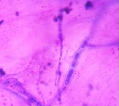
Giemsa stain can also visualize fungi, although it's not the most common method. This staining primarily targets fungal nuclei, revealing their number and location within cellular structures. While not suitable for detailed fungal identification, Giemsa stain serves as a useful tool for preliminary investigations.
Giemsa stain demonstrates good efficacy against many filamentous fungi and yeasts. Preparations can be directly stained or hydrolyzed in N HCl before staining. Staining reagents for yeast chromosomes include 16 drops of Gurr's Giemsa R66 dissolved in 10-12 ml of pH 6.9 Gurr's Giemsa buffer solution.
A blood smear is a thin layer of blood spread on a microscope slide and stained. Giemsa stain distinguishes various components of blood cells by using specific hues to color different parts, allowing for:
The stain highlights the size, shape, and internal structure of red blood cells, white blood cells, and platelets.
Giemsa stain aids in identifying different types of white blood cells including lymphocytes, monocytes, neutrophils, eosinophils, and basophils based on their nuclear shape, chromatin pattern, and cytoplasmic granules.
Here's a detailed account of how Giemsa stain colors different blood cell components:
(1) Red Blood Cells: Stain light pink to red.
(2) Platelets: Appear as small, light pink fragments.
(3) White Blood Cells: Nuclei stained with chromatin appear magenta.
(4) Nuclei: Stain deep purple or magenta.
(5) Cytoplasm: May be light blue (lymphocytes, monocytes) or contain pink or red granules (neutrophils, eosinophils, basophils).
By analyzing Giemsa-stained blood smears for quantities, morphologies, and relative proportions of different cell types, hematologists can diagnose various blood disorders including:
(1) Anemia: Reduced or abnormal red blood cell count.
(2) Leukopenia: Abnormally low white blood cell count.
(3) Leukocytosis: Abnormally high white blood cell count.
(4) Leukemia: Abnormal proliferation of white blood cells.
(5) Infections: Increased counts of specific white blood cells may indicate different types of infections.
Giemsa stain technology finds extensive applications across medical, biological, and scientific research domains. Its role in observing cell structures, studying disease mechanisms, and diagnosing disorders is pivotal. As scientific advancements continue, Giemsa stain technology is poised to further contribute to advancements in human health and life sciences.
[1] https://malariajournal.biomedcentral.com/articles/10.1186/s12936-017-1975-9
[2] https://en.wikipedia.org/wiki/Giemsa_stain
[3] https://microbenotes.com/giemsa-stain-principle-procedure-results-interpretation/
[4] https://www.pnas.org/doi/full/10.1073/pnas.022437999
[5] https://www.ncbi.nlm.nih.gov/pmc/articles/PMC1017366/
[6] Jinan Bibo Biotechnology Co., Ltd. A Giemsa staining solution kit. 2019-10-01.
[7] Guangzhou Dahui Biotechnology Co., Ltd. A chromosome staining method and its application. 2024-02-06.
[8] https://www.sciencedirect.com/topics/biochemistry-genetics-and-molecular-biology/giemsa-stain
 |
 |
|
 |
|
|
 EN
EN
 Agricultural News
 Food News
 Industrial News
 Cosmetic News
 Pharmaceutical News
 Science News