-
We detected your language preference as English. Would you like to switch to the English version for a better experience?
Switch to English
Stay here


Born in Germany in 1867, Gustav Giemsa was primarily involved in chemical work until his death in 1948. The Giemsa stain, named after him, was initially used to demonstrate parasites in malaria but has since been applied in histology due to its high-quality staining of chromatin and nuclear membranes, differential staining of certain cell components, and variability in cytoplasmic staining quality depending on cell type. The use of Azure B and its mixture with Azure A in the form of Eosin Methylene Blue can stabilize the staining and its outcome. Giemsa stain is considered the standard diagnostic technique for malaria parasites worldwide and is fundamental in the classification of lymphomas in the Kiel classification.
Named after a German chemist who applied multiple reagents to demonstrate the existence of malaria parasites, Giemsa stain belongs to a group of Romanovsky stains. These neutral stains consist of a mixture of oxidized methylene blue, Azure, and eosin Y, and they are performed on air-dried smears fixed in methanol. Romanovsky stains are used in the pathological examination of cell differentiation, blood, bone marrow membranes, and parasites such as malaria.
(1) Preparation of Giemsa Stain Solution: Dissolve 3.8 grams of Giemsa powder in 250 ml of methanol, heat to 60°C, then slowly add 250 ml of glycerol to the solution. Filter the solution and let it stand for approximately 1-2 months before use.
(2) Preparation Solution: Add 10 ml of prepared solution to 80 ml of distilled water and 10 ml of methanol.
(3) On clean, dry microscope slides, prepare the specimen into thin slices and air dry.
(4) Immerse the smear (2-3 dips) in pure methanol to fix the smear and air dry for 30 seconds.
(5) Submerge the slide in 5% Giemsa stain solution for 20-30 minutes.
(6) Rinse with tap water and air dry.
(7) Add a thick bloodstain and air dry for 1 hour on a staining rack.
(8) Dip the thick blood smear into a diluted Giemsa staining solution (take 1 ml of the original solution and add to 49 ml of phosphate buffer or distilled water, but the result may vary).
(9) Immerse the smear in buffered water distilled water for 3-5 minutes. Air dry.
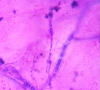
Rotate disease section cut with Giemsa stain:

(1) Advantages:Readily available, easy to prepare, maintain, and use.
(2) Limitations:The working Giemsa stain must be prepared shortly before use.
(1) Safety Guide:Giemsa stain is a common laboratory staining reagent and requires careful handling due to its potential health hazards. When handling, always wear personal protective equipment such as gloves, goggles, and laboratory coats. Avoid inhaling fumes or direct contact with skin and eyes. Ensure good ventilation in the workplace. Store Giemsa stain in sealed containers, away from heat, light, and fire sources. Dispose of waste according to local hazardous materials regulations.
(2) First Aid Measures:If Giemsa stain comes into contact with your skin, wash the affected area thoroughly with soap and water. If it comes into contact with your eyes, open your eyelids and rinse with water for at least 15 minutes. If inhaled, move the patient to fresh air and seek medical attention if necessary. If swallowed, do not induce vomiting. Rinse mouth with water and seek immediate medical attention or poison control center. Remember to have the safety data sheet (SDS) for Giemsa stain ready at all times for detailed first aid instructions.
Giemsa stain technology plays a crucial role in biological research, providing scientists with a reliable method to observe and analyze the structure of cells and tissues. Through this simple yet effective staining method, we gain deeper insights into the microscopic world within organisms, advancing medical diagnostics and scientific research. Hence, the widespread application of Giemsa stain across different fields will continue to drive developments in the life sciences.
 |
 |
 |
 EN
EN
 Agricultural News
 Food News
 Industrial News
 Cosmetic News
 Pharmaceutical News
 Science News